OUR SERVICES
Skin Cancer Screening
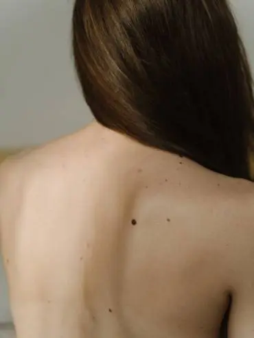
We help you feel confident in your own skin
cancerous and benign skin tumours
Early Cancer Screening Detection
Detecting cancerous and pre-cancerous moles and lesions, skin cancers or melanomas early is paramount for successful treatment and resolution before things get worse. Cancer is a scary prospect, so it is important to check and screen effectively if you have any concerns about marks, lumps, bumps, or moles on your skin.
At Blemish Clinic, we have a trained and experienced dermoscopist who uses dermoscopy equipment to detect potential cancerous tumours, and other significant dermatological skin concerns, that may not be cancerous, but still require removal.
Dermoscopic Screening of Moles and Pre-cancerous Lesions
Both non-pigmented and pigmented skin tumours can be detected with a high accuracy using dermoscopy, compared with simply casting a clinical eye over the skin, or using skin scrapping analysis.
The use of the dermoscopy equipment needs both training and experience in dermatology and dermatoscopy. Blemish Clinic founder, Jan Birch is a dermatology specialist nurse, trained in dermoscopy who has over 40 years’ experience in nursing and over 20 years in dermatological practice. Working alongside a multi-disciplinary team of doctors, dermatologists, and surgical specialists, she offers a comprehensive dermoscopic mole screening medical service for patients wishing to seek out private referral options for cancer checks plus onward skin cancer treatment such as Mohs surgery.
How our patients rate their experience in clinic
*Data gathered via our post-treatment survey
Poor
Average
Excellent
what we do
Skin Cancer Screenings at our Lancashire Clinic
Diagnosis and detection of melanoma and non-melanoma skin cancers including basal cell carcinoma and squamous cell carcinoma. Actinic and seborrheic keratoses are usually not serious however often require checking to rule out anything more sinister.
What Our Clients Say…
“Visited Jan regarding a mole I didn’t think much was wrong with and it turned out to be something much more sinister and if it wasn’t for Jan things could have been much worse, so a big thank you to Jan and her team. They made me feel at ease and have been there all the way couldn’t thank them more.”
Glyn Gilder
FAQs
What is dermoscopy and skin cancer screening?
Dermoscopy (also called dermatoscopy) is a combination of two words – dermis and microscope – which means that it refers to the examination of the skin (the dermis) using skin surface microscopy.
A dermatoscope is a handheld device that looks very similar to a magnifying glass. It is held close to the skin where the concerning lesion is located and allows the medical practitioner to examine the area more closely than with the naked eye. The dermatoscope has high powered lights and magnifies the skin by up to 10 times, so you can see the differing colours and structures within the lesion to aid diagnosis.
Dermoscopy is used to screen moles and lesions for markers that indicate cancerous tissue – this may include the symmetry or asymmetry of the lesion, how uniform it is, the definition of the edges, the distribution of pigment or differing colours within, and other considerations like blood supply, ulceration, skin flaking, cracks etc. By having a close-up view with a dermatoscope, we can more accurately determine if a lesion is benign, pre-cancerous, or cancerous in nature thanks also to well catalogued medical definitions for different presentations of lesion types.
What are the different types of pigmented lesions that can be skin cancer?
Using dermoscopy to look at the pattern of pigment within a lesion we can help to identify and diagnose the lesion, as well as the potential for it to be benign (non-harmful) or malignant and cancerous.
Here are some short explanations of terms that you may hear.
Lentigos/Lentigines (freckles)
Freckles (ephelides) are a common occurrence and inherited characteristic, often in fair-skinned individuals, and especially in children and adults with red hair, where it is believed to be genetically linked. These are harmless and such individuals usually just need regular and high factor sun protection. The profusion of freckles may become more, or less obvious between summer and winter.
Lentigines or lentigos look very much like freckles but are caused by sun exposure or UV light from sunbeds – often meaning they are referred to as solar lentigines. Again, more common in fair skinned individuals, they also become very common in people after the age of 40 due to their lifetime exposure to the sun. Solar lentigos are brown, flat marks that are more defined than usual freckles, and do not fade in the winter. Solar lentigos can evolve into one or more seborrheic keratoses and do have the capacity to become malignant (lentigo maligna) so should be checked as they can be difficult to differentiate from a benign lentigo.
Seborrheic keratosis (age spots)
A seborrheic keratosis is a common skin growth seen as people get older, they are often brown, tanned, or black in colour, raised or flat, and called age spots or less kindly, senile warts. Seborrheic keratoses are benign and noncancerous; their cause is unknown, but they can come from a solar lentigo. Often people develop several.
Although they are not associated with being precancerous, they can be difficult to tell apart from a skin cancer such as a melanoma, basal cell carcinoma, or squamous cell carcinoma, so should be checked.
Actinic keratosis
An actinic or solar keratosis is caused by sun damage and is a dry, scaly patch of skin, that may be itchy. They are usually benign, but can be precancerous so it is important to have them checked, especially if the lesion has altered in any way recently – started bleeding, increased in size, changed colour etc.
Naevus (moles)
A mole is a melanocytic naevus that is a common and benign, or harmless skin lesion. They can appear in babies and children, even being present at birth (congenital) or be acquired later in life, often related to sun exposure. Most of us will have at least one mole, but usually can pinpoint several located in various places on the body. Those with fair skin tend to have more moles. They come in all shapes and sizes, mostly round or oval, raised or flat, varying in colour, and are categorised accordingly – junctional (flat mole), dermal (raised nodule), compound (a raised area on top of a flat patch), or combined (two distinct types of moles within the same lesion).
If the size, shape, or colour of a mole changes, or it starts to itch, crust, or bleed, or a new mole suddenly appears and you are over 40, it is important to have it checked as it may be evolving into a malignant melanoma.
Most moles are harmless, but you may wish to have them removed as a cancer prevention, or if they are in a location that causes irritation with clothing or jewellery, or when shaving for men, or if you feel that they are cosmetically unsightly. See our page on mole removal.
Basal cell carcinoma
A basal cell carcinoma (BCC) is a non-melanoma skin cancer. It is a type of locally invasive skin cancer, also called a rodent ulcer that develops in the basal cells found at the bottom of the epidermis, the outermost layer of the skin. Basal cells are responsible for producing new skin cells. BCC is the most common type of skin cancer.
This type of skin cancer is more common in elderly men but can also affect younger people and women. Sun exposure history including repeated sun burn, fair skin and red hair are all predispositions for BCC formation. BCCs come in various types but a nodular or lumpy lesion, often with a small central ulceration or crater (hence the name rodent ulcer) is the most common. Because they are a local skin cancer, they are rarely a threat to life, but should be checked and treated.
Squamous cell carcinoma
A (cutaneous) squamous cell carcinoma (SCC) is the second most common type of skin cancer. It is a non-melanoma skin cancer also called a keratinocyte cancer because it is formed from the cells in the skin that make keratin for skin, hair, and nails. SCCs develop in the squamous cells in the middle and outer layers of the skin. It is an invasive skin cancer, meaning that it can spread, but is usually not life-threatening unless left untreated.
This type of skin cancer is common in the same people as other forms of skin cancer, but the risk of developing an SCC is greater in those with a previous BCC or melanoma. Sun exposure, an outdoor occupation, smoking, and certain medications to treat autoimmune diseases can increase the risk of developing an SCC.
SCCs tend to be scaly or crusty in appearance, often tender or painful, and evolve from actinic keratoses. Such lesions should be checked and treated swiftly to avoid further complications.
Melanoma
Melanoma or malignant melanoma is the third most common type of skin cancer and is very serious. The charity Melanoma UK notes that around 2,500 people die from malignant melanoma every year in the UK, so it is vital to get any suspicious moles checked immediately.
Melanoma is an uncontrolled growth in melanocytes which are the pigment cells within the skin that produce melanin (the response that makes us tan to protect our skin by absorbing UV radiation from the sun). As humans, we all have the same amount of melanocytes, but those with darker skin types (black and brown) produce more melanin than fairer, white skin types so they are less likely to experience skin damage from UV radiation.
Non-cancerous growths in these melanocyte cells are what produce freckles (ephelides and lentigines) and moles (benign melanocytic naevi). If a cancerous growth occurs in the melanocytes, arising from normal-looking skin or through changes in a freckle or mole, this is a melanoma. These can be localised, but can also be invasive, spreading in the skin, or to other parts of the body that can prove fatal if not discovered soon enough.
Unlike other skin cancers that can be more common in the very elderly, malignant melanoma tends to occur in a younger population from their 30s through to their 60s. The main environmental cause of melanoma is sun exposure, with increased risk within populations in areas of high solar radiation and with fair skin types, including Europe, American, Australia and New Zealand. A high rate of sunbathing, tanning and sunburn incidents, fair skin, high density of freckles, red or blonde hair, blue or green eyes, and multiple moles, including multiple atypical moles (that appear different to the others) make a person more at risk of melanoma. The presence of non-melanoma skin cancers like actinic keratoses approximately doubles the risk of developing a melanoma.
This type of skin cancer is often regarded as the most common in men after prostate and colorectal cancers, and the most common in women after breast and colorectal cancers. A previous history of melanoma also increases the risk of developing future melanomas.
The first sign of a malignant melanoma is when an existing mole or freckle starts to change; it is important to detect them early, so if you have noticed any changes your moles should be checked.
How to check if a mole may become cancerous
A mole or melanocytic naevus is a common skin lesion that is caused by a proliferation or rapid increase in melanocytes (pigment creating cells) within the skin. Moles are usually harmless and can be congenital from birth or acquired as we develop and age.
If a mole has unusual features or is “funny-looking” it may be an atypical (melanocytic) naevus. This is also called the “Ugly Duckling” sign and is where a mole looks different to most other moles present on an individual. These are usually the type of mole that warrant further inspection and checking as they can be suspected of being a melanoma or cancerous mole.
An atypical naevus may be large (over 5mm in diameter), have poorly defined edges, and be irregular in shape with varying colours including pink, tan, brown, and black, and be made up of flat and bumpy areas.
When screening moles for suspected cancerous characteristics, we use the ABCDE rule of skin cancer. This is also a useful tool for you to use to determine if you need to make an appointment to come and see us for mole screening. If you have two or more of these characteristics it would warrant rapid skin cancer screening with our specialist team.
A – Asymmetry: a mole with an asymmetrical shape or pigmentation pattern
B – Border: a mole with an irregular or uneven border, often described as a geographical edge, rather than simply round or oval
C – Colour: a mole with variable colours within, usually two or more
D – Diameter: a mole with a diameter greater than 5mm
E – Evolution: a mole that is showing signs of change over time
Recently this rule has been extended as follows for additional characteristics:
E – Elevation: a mole that is becoming raised
F – Firm: a mole that feels firm or solid to the touch
G – Growing: a mole that is showing signs of change over time
Bleeding, crusting, or itching are also signs of concern.
If you suspect you have a mole that is showing two or more of these characteristics, please book a consultation with us at the Blemish Clinic. It may be nothing, but skin cancers benefit from early detection and rapid treatment. Using dermoscopy, we can get a close-up look at the ABCDE characteristics to increase our ability to diagnose if the mole is indeed malignant.
Guideline recommendations by NICE (The National Institute for Health and Care Excellence) advise the use dermoscopy in the assessment of pigmented skin lesions by trained healthcare professionals. Their rationale states that, “Dermoscopy performed by suitably trained specialists is more sensitive and more specific in classifying skin lesions than clinical examination with the naked eye. It lessens the chance of missing a diagnosis of melanoma and reduces the number of unnecessary surgical procedures to remove benign lesions”.
Am I suitable for skin cancer screening and how do I access the service?
At Blemish Clinic, we treat non-melanoma skin cancers and photodamage using topical treatments, Photodynamic Therapy (PDT), cryotherapy, or surgical excision where required.
Our team collaborate and work as part of a multi-disciplinary team (MDT) working across oncology, plastics, and Mohs surgery. Mohs surgery is a microscopically controlled surgical technique used to treat skin cancer where layers of skin are removed progressively, and each layer is examined under a microscope until a layer of tissue is reached that is cancer-free.
Your initial consultation will be with either our dermatology specialist nurse, Jan Birch or consultant dermatologist, Dr Andrew Winter, where your mole or skin lesion will be accessed visually and using dermoscopy to diagnose and decide on the most appropriate treatment option, which may include further referral within the MDT.
A full medical history will be taken to determine if there are any underlying health issues, medications, or past skin cancer history which could be impacting on the formation of your skin lesions, or conflict with any proposed treatments, alongside any known allergies.
Following assessment and diagnosis, which may also include a biopsy, a treatment plan and referral journey can be developed. If your lesion is a non-melanoma cancer, we will offer treatment options in clinic, melanomas and more complex cancerous lesions will be referred to our Mohs surgeon.
Blemish Clinic provide easy access to continued care which may not be available through primary healthcare services. We have a holistic approach to practice, providing continuity of care on your treatment journey through out our MDT pathway, which includes help with wound healing, and optimising both your medical and cosmetic outcome from dermatological and skin cancer treatments.
What should I expect from skin cancer screening?
At Blemish Clinic, our speciality is dermoscopy, which allows us to look deeper into the skin to diagnose skin lesions. Our specialist nurse Jan Birch and Dermatology Consultant will use their clinical skills combined with their dermatoscopic skills, the two are very different but are essential for determining an accurate diagnosis of the many pigmented and non-pigmented lesions, both benign and malignant. Blemish Clinic works alongside a multidisciplinary team and will provide an onward referral to ensure you have access to both rapid and urgent screening appointments and ongoing treatment if required.
How much should my treatment cost?
We believe in being transparent with our prices and want you to be fully aware and comfortable with the cost of your treatment. A full list of our prices can be found here. We will always agree a final price with you though before treatment commences.
To assess and diagnose pigmented and non-pigmented lesions, including moles, for the presence of skin cancers such as melanoma or non-melanoma skin cancers – basal cell carcinoma, squamous cell carcinoma, actinic and seborrheic keratoses. Determination of onward treatment can be planned following diagnostic screening for skin cancer.
Using dermoscopy as a high accuracy method of screening lesions and moles for skin cancer, we can avoid missing potential cancerous lesions through naked eye examination alone and remove the need for unnecessary biopsies (skin scrapping) in the diagnosis of melanoma, cancerous, and pre-cancerous lesions.
NICE guidelines recommend the use of dermoscopy in skin cancer screening and the assessment of pigmented skin lesions as a more sensitive and more specific aid to classifying skin lesions than clinical examination with the naked eye. This method of skin cancer screening is highly accurate.
A consultation for skin cancer screening, including assessment by dermoscopy will take one hour approximately.
Skin cancer screening uses diagnostic tools that are not painful or uncomfortable and are designed to assist the practitioner with a close-up view of the lesion or mole of concern.
There is no recovery time needed from skin cancer screening appointments.
AWARDS


